Notice: The following article is for educational and informational purposes only. The substances described are intended for research and laboratory use. This does not constitute medical advice or encouragement to use these substances in humans or animals.
Introduction
TB-500 is a synthetic peptide corresponding to the active domain of Thymosin β4 (Tβ4) — a naturally occurring protein of ~4963 Da that is one of the most abundantly expressed peptides in eukaryotic cells. Thymosin β4 was first isolated from calf thymus by Allan Goldstein in 1981, though it was later demonstrated that its expression is not restricted to thymic tissue — it occurs in virtually all nucleated cell types.
TB-500 replicates the 43-amino acid sequence of Thymosin β4 (Ac-SDKPDMAEIEKFDKSKLKKTETQEKNPLPSKETIEQEKQAGES), containing the key G-actin-binding motif — the LKKTET sequence (positions 17–22). This motif is responsible for the peptide’s biological activity in the context of actin cytoskeleton reorganization.
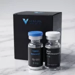
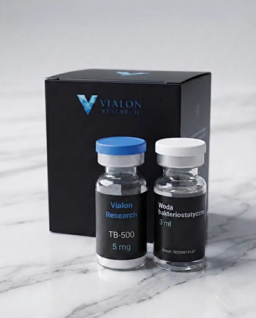
Our shop offers research-grade TB-500 as a lyophilized powder with purity ≥98% HPLC, as well as a TB-500 + BPC-157 blend combining two regenerative peptides with complementary mechanisms of action.
Structure of Thymosin β4 and the TB-500 Peptide
Thymosin β4 is a small, acidic 43-amino acid peptide with N-terminal acetylation (Ac-Ser) and a C-terminal serine residue. Molecular weight is ~4963.4 Da, CAS number: 77591-33-4. Unlike most proteins of this mass, Thymosin β4 is an intrinsically disordered protein (IDP) — it does not adopt a stable tertiary structure in aqueous solution.
This disordered structure is functionally significant — it allows Thymosin β4 to bind different protein partners depending on the cellular context. Key structural motifs include:
- LKKTET motif (positions 17–22) — a sequence that binds monomeric G-actin with high affinity. It forms a short α-helix upon contact with actin, sequestering monomers and inhibiting spontaneous polymerization
- N-terminal domain (1–14) — responsible for interactions with integrins and extracellular matrix proteins
- C-terminal domain (30–43) — a regulatory region subject to post-translational modification
- Methionine residue (Met⁶) — susceptible to oxidation; the primary site of oxidative degradation of the peptide
The presence of methionine at position 6 means that TB-500 is more susceptible to oxidative degradation than, for example, BPC-157, which contains neither methionine nor cysteine. This has direct implications for storage conditions — details are covered in the section below.
Primary Mechanism: G-Actin Sequestration
Actin is one of the most abundant proteins in eukaryotic cells, accounting for up to 10% of total protein mass. It exists in two forms:
- G-actin (globular) — the monomeric form, dissolved in the cytoplasm
- F-actin (filamentous) — the polymerized form, building the microfilaments of the cytoskeleton
The equilibrium between G- and F-actin is dynamic and tightly regulated — it governs cell shape, migratory capacity, division, and interaction with the surrounding environment. Thymosin β4 serves as the primary G-actin buffer in cells, binding monomers in a 1:1 complex and preventing their spontaneous polymerization.
Binding mechanism:
- The LKKTET motif recognizes the binding cleft on the surface of G-actin
- Thymosin β4 forms a short α-helix around actin, blocking binding sites for profilin and other polymerization-promoting proteins
- The Tβ4–G-actin complex remains dissolved in the cytoplasm as a „reserve” of monomers
- Upon cellular signal (e.g., chemokine, growth factor), Thymosin β4 releases G-actin, enabling controlled polymerization and formation of migratory structures (lamellipodia, filopodia)
This mechanism explains why Thymosin β4 is so important in processes requiring cytoskeletal reorganization — cell migration, wound healing, new blood vessel formation, and tissue repair.
Research Areas
Angiogenesis and Blood Vessels
Thymosin β4 promotes angiogenesis — the formation of new blood vessels from existing ones — by stimulating the migration and proliferation of endothelial cells. In vitro models (Matrigel assays) and in vivo models (murine) have demonstrated increased vascular density following Tβ4 administration. The mechanism involves activation of αvβ5 integrins and upregulation of VEGF.
The pro-angiogenic properties of Thymosin β4 distinguish it from many other research peptides and have implications in ischemic models, where collateral vessel formation is critical for tissue survival.
Wound Healing and Tissue Regeneration
In animal models, Thymosin β4 accelerates healing of dermal, corneal, and cardiomyocyte wounds. A key role is played by promoting cellular migration — of fibroblasts, keratinocytes, and endothelial cells — to the site of injury. Additionally, Tβ4 inhibits apoptosis and modulates the inflammatory response, limiting excessive inflammation during the early phase of healing.
These regenerative properties place TB-500 in the same research context as BPC-157 and GHK-Cu — however, the mechanism of each peptide is distinct. TB-500 acts through the actin cytoskeleton, BPC-157 through the NO/VEGF pathway, and GHK-Cu through copper chelation and gene expression modulation.
Cardioprotection
Studies by Bock-Marquette et al. (2004) demonstrated that Thymosin β4 activates Akt kinase (PI3K/Akt pathway) in cardiomyocytes, promoting cell survival following ischemia. In a murine myocardial infarction model, Tβ4 administration reduced infarct size and improved contractile function of the myocardium. Activation of cardiac progenitor cells was also observed — an effect attributed to the ability of Tβ4 to mobilize stem cells from their niche.
Neuroprotection and Remyelination
Growing interest surrounds the neuroprotective potential of Thymosin β4. In spinal cord injury and hypoxic-ischemic encephalopathy models, Tβ4 promoted survival of neurons and oligodendrocytes. Particularly noteworthy are observations regarding remyelination — in demyelination models, Tβ4 stimulated differentiation of oligodendrocyte precursors (OPCs) into mature myelinating cells.
TB-500 vs. BPC-157 — Comparison
TB-500 and BPC-157 are the two most frequently compared regenerative peptides. Despite being classified in the same category, they differ fundamentally:
| Parameter | TB-500 | BPC-157 |
|---|---|---|
| Amino acids | 43 | 15 |
| Molecular weight | ~4963 Da | ~1419 Da |
| Origin | Thymosin β4 fragment (thymus) | Fragment of gastric protective protein |
| Primary mechanism | G-actin sequestration, cytoskeletal reorganization | NO pathway modulation, VEGF/EGF |
| pH stability | Neutral (7–8) | Stable at pH 2–10 |
| Oxidation susceptibility | Yes (Met⁶) | No (no Met/Cys) |
| CAS | 77591-33-4 | 137525-51-0 |
The complementarity of mechanisms — actin cytoskeleton (TB-500) vs. angiogenesis/NO (BPC-157) — is the reason both peptides are often studied together. The ready-made TB-500 + BPC-157 blend in a 1:1 ratio eliminates the need to prepare two separate solutions. For more on the differences between these peptides, see the comparison section on the TB-500 + BPC-157 product page.
Storage and Stability of TB-500
TB-500 requires more careful storage than some other peptides due to the presence of the methionine residue (Met⁶) that is susceptible to oxidation:
- Lyophilized powder: store at ≤ −20°C, under argon or nitrogen atmosphere if possible. Protect from light and moisture. Stability exceeds 24 months under these conditions.
- After reconstitution: solution in bacteriostatic water should be stored at 2–8°C and used within 14 days. Avoid repeated freeze-thaw cycles.
- Degradation: the primary degradation pathway is oxidation of Met⁶ to methionine sulfoxide, which reduces affinity for G-actin. UV light exposure and elevated temperature accelerate this process.
For further reading on peptide degradation mechanisms and storage best practices, see our knowledge base articles on peptide stability and our guide on peptide storage.
Frequently Asked Questions
What is TB-500?
TB-500 is a synthetic 43-amino acid peptide corresponding to Thymosin β4 — a naturally occurring protein first identified in the thymus. It contains the LKKTET motif that binds G-actin. Molecular weight ~4963 Da, CAS: 77591-33-4.
What is Thymosin Beta-4?
Thymosin β4 (Tβ4) is an endogenous protein present in virtually all nucleated cells. It serves as the primary G-actin buffer — binding actin monomers and regulating cytoskeletal dynamics. TB-500 is a synthetic counterpart used in research.
How does TB-500 differ from BPC-157?
TB-500 (43 aa, ~4963 Da) acts by binding G-actin and reorganizing the cytoskeleton. BPC-157 (15 aa, ~1419 Da) modulates the nitric oxide pathway and growth factors VEGF/EGF. They have complementary mechanisms — also available as a TB-500 + BPC-157 blend.
What is the primary mechanism of action of TB-500?
TB-500 binds monomeric G-actin through the LKKTET motif (positions 17–22), sequestering monomers and regulating the polymerization of actin filaments (F-actin). This mechanism is central to cell migration, wound healing, and angiogenesis.
How should TB-500 be stored?
Lyophilized powder should be stored at ≤ −20°C (stability >24 months). After reconstitution in bacteriostatic water, store at 2–8°C and use within 14 days. TB-500 contains methionine susceptible to oxidation — protect from UV light and store in sealed vials.
Bibliography
- Goldstein AL et al. (1966). „Purification and biological activity of thymosin, a hormone of the thymus gland.” Proc Natl Acad Sci USA, 56(3):1010–1017.
- Safer D, Elzinga M, Nachmias VT (1991). „Thymosin beta 4 and Fx, an actin-sequestering peptide, are indistinguishable.” J Biol Chem, 266(7):4029–4032.
- Bock-Marquette I et al. (2004). „Thymosin β4 activates integrin-linked kinase and promotes cardiac cell migration, survival and cardiac repair.” Nature, 432(7016):466–472. doi:10.1038/nature03000
- Malinda KM et al. (1999). „Thymosin beta4 accelerates wound healing.” J Invest Dermatol, 113(3):364–368. doi:10.1046/j.1523-1747.1999.00708.x
- Philp D et al. (2004). „Thymosin beta4 promotes angiogenesis, wound healing, and hair follicle development.” Mech Ageing Dev, 125(2):113–115. doi:10.1016/j.mad.2003.11.005
- Morris DC et al. (2010). „Thymosin β4 improves functional neurological outcome in a rat model of embolic stroke.” Neuroscience, 169(2):674–682. doi:10.1016/j.neuroscience.2010.05.017
- Sosne G et al. (2007). „Thymosin beta 4 promotes corneal wound healing and modulates inflammatory mediators in vivo.” Exp Eye Res, 85(5):620–628. doi:10.1016/j.exer.2007.07.013